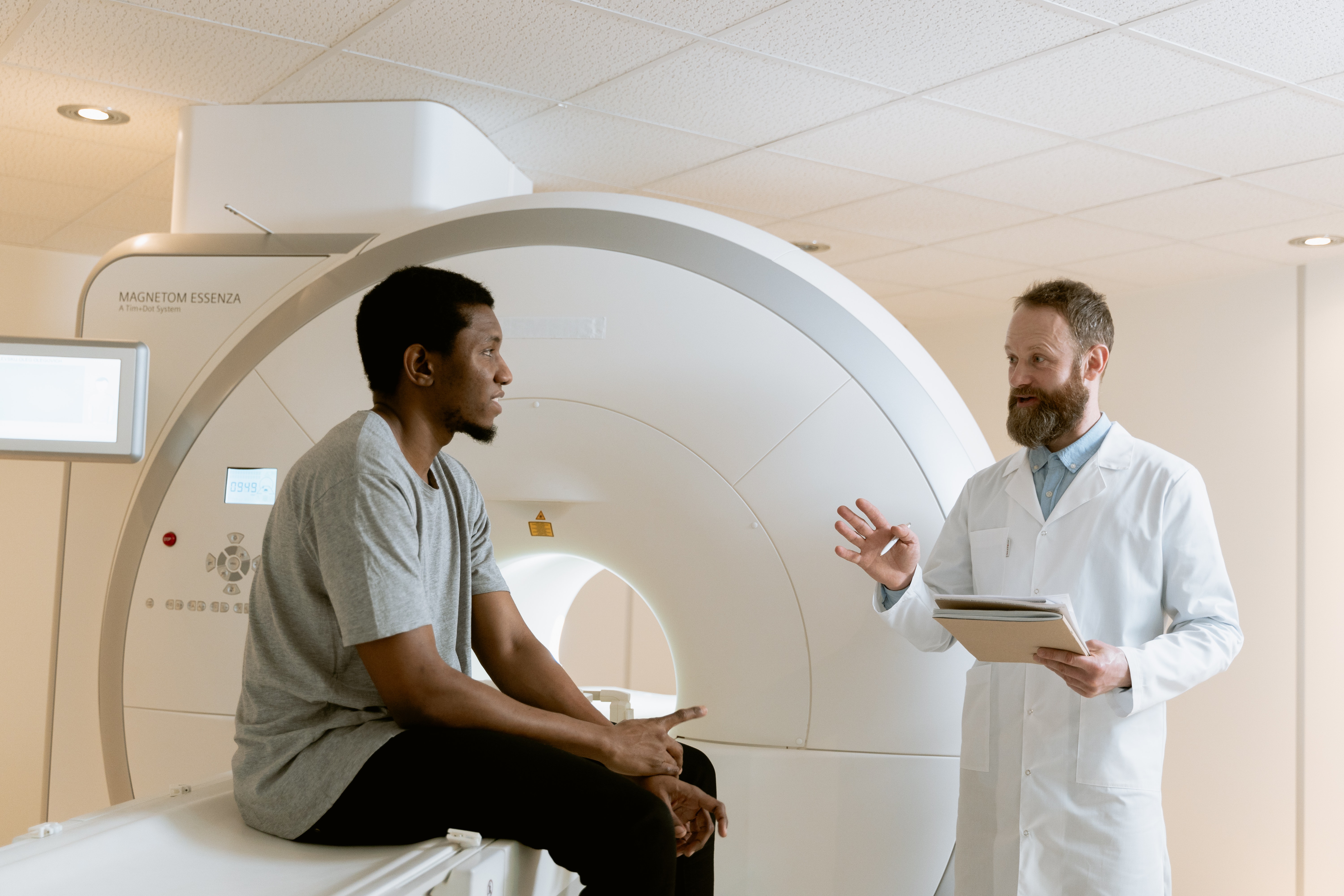
This research theme is interested in peripheral and central pain mechanisms, meaning what happens in the nerves and in the brain. We explore these mechanisms through different types of imaging techniques (scans), as well as a technique called microneurography, which involves reading electrical signals directly from small nerve fibres.
Peripheral nerve imaging
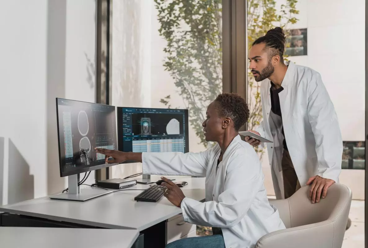
Magnetic resonance imaging (MRI) can reveal detailed structural properties of nerves. It has shown that diabetic and entrapment neuropathies cause changes in these structural properties, which correlate with sensory function and physiology. The PAINSTORM teams at Imperial College London and the University of Oxford have been pioneering peripheral nerve imaging at both 3 and 7 Tesla (these are units describing the strength of the magnets in a scanner), which will be used to determine structural phenotypes in neuropathic pain. We have developed sciatic nerve imaging at Imperial College London and median nerve imaging at the University of Oxford. We will study these imaging metrics in relation with other measures in well-phenotyped patients with different types of neuropathies.
Microneurography
Microneurography is the only neurophysiological technique that records neuronal activity directly from nociceptors in awake patients. We must understand better how changes in peripheral nociceptive fibre properties in disease states (e.g. diabetes) contribute to neuropathic pain. With deep phenotyping, microneurography, and peripheral nerve imaging, we will be able to compare the excitability profile of each nociceptive fibre subclass and their responses to external stimuli between study participants with painful and painless diabetic polyneuropathy.
We will also look at how changes in nociceptive fibre properties correlate with clinical variables, such as neuropathy severity or diabetic control, and peripheral nerve imaging metrics. We will stratify patients into sensory profiles to determine whether these correlate with microneurography.
Spinal cord, brainstem, and whole brain neuroimaging
Neuropathic pain involves central and peripheral mechanisms. A central theme to our work has been to prove that an endogenous dysfunctional descending pain modulatory system is a key pathophysiological mechanism in chronic pain. Scaling up this observation and better understanding what drives this dysfunction is the next key goal. This will be enabled by linking to Work Package 3 outcomes.
We have built on early work in learning/prediction errors in pain to show similar learning-related effects in the spinal cord. Building on our track record of neuroimaging innovation, we will take subsets of patients to further verify DRG-dorsal horn functional connectivity and dorsal horn activity as metrics of aberrant peripheral nerve activity and dorsal horn sensitisation, respectively.
We will collaborate with Work Packages 2 and 3 to better determine imaging endophenotypes of these psychosocial processes that powerfully contribute to the development, exacerbation and maintenance of persistent pain.
Microneurography
In Oxford, we used microneurography, an in vivo technique that records activity from single human peripheral nerve fibres, to investigate how peripheral neuronal hyperexcitability drives neuropathic pain. We recruited patients with diabetic and non-diabetic polyneuropathies, including small fibre neuropathy and other rare neuropathic pain disorders, alongside healthy controls. 253 participants were recruited and underwent deep phenotyping combining clinical assessment, histology, quantitative sensory testing, imaging, biomarker analysis, nerve conduction studies, and microneurography. A total of 188 successful recordings were obtained, a 74.3% success rate that has allowed us to build one of the largest microneurography datasets in neuropathic pain research. Through this approach, we aim to identify abnormal activity in nociceptors and determine how peripheral neuronal dysfunction shapes different neuropathic pain phenotypes, ultimately improving patient stratification and supporting the development of precision therapies.
We are also completing a systematic review in which we evaluate microneurography methodologies, describe current approaches to classifying C fibres, and synthesise reported abnormalities in peripheral nerve activity across neuropathic pain conditions
Peripheral Nerve imaging
We have developed novel imaging approaches using 7T MRI to better understand the structural properties associated with peripheral neuropathy and how these correlate with sensory function. We have carried out a pilot study and recruited a cohort of 24 people with a range of peripheral neuropathies (diabetic, HIV, small fibre) plus healthy controls, who have undergone peripheral nerve imaging, combined with detailed pain phenotyping, harmonsing our assessment with that used in Oxford and Dundee. This work demonstrated proof on concept that it was possible to generate very detailed, high-resolution images of the sciatic nerve at the level of the thigh in patients and healthy volunteers. It also demonstrated the challenge of scanning at high field strength, with significant numbers of patients (particularly those with diabetes and HIV) being excluded due to metalwork in their bodies, including cardiac and kidney stents, and joint replacements.
We plan to publish this work to showcase this imaging technique, comparing healthy controls with neuropathy groups, looking at the size of the nerves, as well as how “bright” they are on specific types of MRI sequence. We have also published a systematic review of MRI findings in diabetic and HIV-associated neuropathy in preparation for this work, and edited a special edition on Peripheral Nerve Imaging for Frontiers in Neuroscience.
Brain imaging
Two centres (Oxford and Dundee) carried out brain imaging to better understand central mechanisms of neuropathic pain, and why some people with nerve damage develop severe, persistent pain while others do not.
- In Oxford, we focussed on diabetic neuropathy, establishing a deep-phenotyping brain imaging approach, combining fMRI with detailed clinical assessment and microneurography to study pain at the level of the brain and the whole person. Analysis is ongoing to identify the different mechanisms driving pain in painful diabetic peripheral neuropathy — a first step towards matching treatments to the mechanism causing an individual's pain.
- Brain imaging completed in 30 patients with diabetic neuropathy, spanning painful and painless forms, scanned and deeply characterised, generating the brain-connectivity measures behind the work package's questions about the brain's descending pain control system.
- In Dundee, we focussed on people undergoing neurotoxic chemotherapy as part of their cancer treatment. We have collected structural and functional imaging on people both before and after completion of chemotherapy, plus healthy controls, that will allow us to better understand risks of developing chemotherapy-induced peripheral neuropathy (CIPN).
- Current analysis are ongoing to understand whether chemotherapy alters reward-learning behaviour and its neural signature, and critically whether reward-learning measured before chemotherapy can predict who goes on to develop chemotherapy-induced peripheral neuropathy (CIPN). Analyses compared a pre-chemotherapy group (n = 18) with a post-chemotherapy group (n = 39).
- Preliminary findings: CIPN exposure was associated with slower stimulus processing but quicker evidence accumulation, with a reduction in overall decision time in a win-loss reward task. More severe pain was linked to blunted reward prediction-error responses but better option discrimination. Reward- and loss-outcome signals were blunted after chemotherapy across pain- and salience-relevant regions - nucleus accumbens and insula (reward); periaqueductal gray, insula, posterior cingulate, dorsolateral prefrontal cortex and hypothalamus (loss). These brain-level changes mirror the behavioural findings. As behaviour was measured before pain onset, these are candidate early predictors of CIPN risk.
- The results suggest that pre-chemotherapy behavioural and imaging measures may facilitate estimation of risk of developing CIPN. Convergent behavioural, computational and neural evidence suggests chemotherapy dampens outcome processing, and that normal pre-chemotherapy variation in reward learning may flag who is at risk of CIPN.
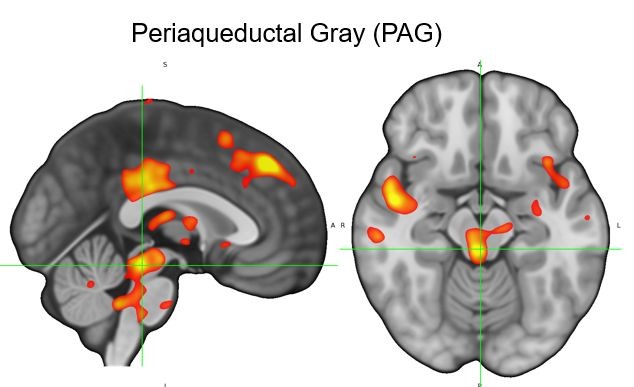
This figure shows increased activity in the periaqueductal grey when comparing scans after chemotherapy with before.
Patient involvement
- Co-production of workshops for the 9th International Congress on Neuropathic Pain, Lisbon, 2023:
- Workshop Presentations: Patient and Public Involvement (PPI) in Neuropathic Pain Research: What It Is (and is Not), Why It Matters, and How to Build Confidence and Capacity
- Chemotherapy Induced Peripheral Neuropathy: Challenges and Progress
Co-production of workshop for the 10th International Congress on Neuropathic Pain, Berlin, 2025: It Takes a Community: Approaches to Understand and Prevent Chemotherapy-Induced Peripheral Neuropathy
- People living with chronic pain, who had experience of being participants in an earlier study (PiNS study) took part in one-to-one interviews to help shape the Oxford imaging protocol. Their input directly shortened the scanning session from nearly two hours to around 45 minutes, and replaced a prolonged (tonic) heat stimulus with a briefer, more tolerable one.
Understanding Chronic Pain and advances in research – NIHR article, 23/01/2025
- Kelleher EM, Lange F, Wanigasekera V, Rathod-Mistry T, Nichols T, Seymour B, Tracey I, Segerdahl AR, Irani A. Brain signatures of nociplastic pain: Fibromyalgia Index and descending modulation at population level. Brain. 2026;149(4):1365–1380. doi:10.1093/brain/awaf307.
- Segerdahl AR, Deli A, Raghu ALB, Crockett R, Denison T, Green AL, Tracey I. Precision-guided multi-site insular deep brain stimulation for refractory neuropathic pain. Brain Stimulation. 2026;19(1):102999. doi:10.1016/j.brs.2025.102999
- Themistocleous AC, Baskozos G, Bennet DLH, Serra J. Defining the link between peripheral neuronal activity and neuropathic pain: observational study protocol to investigate in vivo neurophysiological properties of nociceptors in patients with chronic neuropathic pain. PONE-D-26-06203R1 10.1371/journal.pone.0349407
- John Locke, Goknur Selen Kocak, David L.H. Bennett, Jordi Serra, Cassie Higgins, Andreas Constantinos Themistocleous. Microneurography techniques for recording and characterising C fibres in humans and non-human primates: a systematic review protocol. PROSPERO 2026 CRD420261346067. Available from https://www.crd.york.ac.uk/PROSPERO/view/CRD420261346067.
- Systemic anti-cancer therapy associated with the occurrence of peripheral neurotoxicity and, specifically, peripheral neuropathy. Higgins C, Gauthier LR, Smith BH, Colvin L. Int J Cancer. 2026; 159(2): 460-466. doi: 10.1002/ijc.70414
- Magnetic resonance imaging as a biomarker in diabetic and HIV-associated peripheral neuropathy: a systematic review-based narrative. Evans, MC, Wade C, Hohenschurz-Schmidt D et al. Front Neuro 2021; 15:727311. doi: 10.3389/fnins.2021.727311